08/24/2007
Analysis of
subcellular structures isolated from single cells
Recent Research from the group of Professor
Edgar Arriaga.
In order
to maintain cellular function, subcellular structures (organelles) have
a complex organization and a wide range of properties even for organelles
of the same type. In particular, the architecture of mitochondria varies
from a tight network to hundreds of independent organelles, which collectively
fulfill mitochondrial roles (e.g. ATP production, lipid metabolism, and
calcium homeostasis) in the cell. Because organelle-to-organelle variations
are of high importance in biological processes such as embryogenesis, tissue
differentiation, and disease, there is great demand to develop highly sensitive
techniques to investigate the properties of organelle ensembles in single
cells. In the January issue of Analytical and Bioanalytical Chemistry (Anal. Bioanal. Chem. 387, 107-118,
2007), Ryan D. Johnson and co-workers in the Arriaga group reported the
first analysis of mitochondria isolated from single 143B human osteosarcoma
cells. These cells were labeled via expression of the fluorescent protein
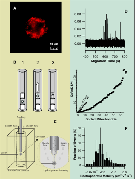
DsRed2, targeted to mitochondria (Figure 1A). Subsequently, a single cell
was introduced into the lumen of a capillary where the organelles are released
by a combined treatment of digitonin and trypsin (Figure 1B). After this
treatment, an electric field was applied to cause electromigration of the
released organelles toward a laser-induced fluorescence detector (Figure
1C). From an electropherogram (Figure 1D), the number of detected events
per cell, their individual electrophoretic mobilities (Figure 1F), and
their individual fluorescence intensities (Figure 1E) are calculated. The
Arriaga group is currently working toward extending this new bioanalytical
technology to investigate how giant and normal mitochondria within the
same cell contribute to the aging process and how acidic organelles contribute
to drug resistance.

Figure 1. Analysis of
organelles taken from single cells. (A) A single 143B cell expressing DsRed2,
targeted to mitochondria. (B) One
cell is introduced into a fused silica capillary and placed between two
plugs of buffer capable of disrupting the plasma membrane and the cytoskeletal
network (1), the cell is electrokinetically displaced (2), and upon plasma
membrane disruption, organelles are released (3). (C) Detail
of a post-column laser-induced fluorescence detector. (D) Electropherogram displaying detection
of the individual organelles from a single cell. (E) Intensities and number of organelles
taken from three single cells. (F) Average
of electrophoretic mobility distributions of organelles taken from three
single cells.
|